Tomographic Imaging
Stellarray has designed and prototyped portable tomographic imaging systems using digitally addressable x-ray sources (DAXS) to bring 3-D radiography for cancer screening and other types of medical imaging to new populations and settings. The sources are “stationary” in that the x-ray spot location is addressed electronically without moving a tube (all the spots are inside a common vacuum source). The systems are “portable” because once the usual metal gantry in conventional CT or tomosynthesis systems is gone, the DAXS, a flat panel x-ray detector and structural supports weigh under 15 kG and can fit into a suitcase. By portable we mean the 3-D x-ray imaging system can be carried by one person, so it can be brought to the point of injury or treatment, into the home, onto vehicles and out to underserved populations. Systems that require wheels to move are merely mobile. Tomosynthesis is an emerging radiographic imaging modality in which x-ray projections are taken over a limited angular range instead of the full 360°arc in CT. It is best known for digital breast tomosynthesis (“3-D mammograms”) but it can be used for many other imaging conditions. Stellarray’s sources can paired with detectors in a modular approach to build a variety of system configurations, ranging from one source and one detector, to two sources and one detector, to multiple source-detector pairs. The preferred design has the high voltage power and control system integrated into the same module as the source, to avoid the need for cumbersome and hazardous high voltage cables. Stellarray’s hardware can be the platform for an open system architecture in which other groups build their own reconstruction algorithms or AI systems with Stellarray modules. It is the perfect companion for extending radiographic AI to the edge.
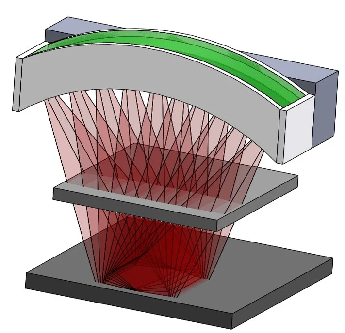
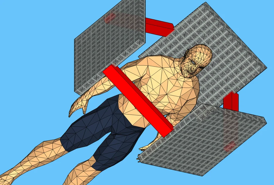
Stellarray completed an SBIR project for NASA in which some of these elements were designed and prototyped. The objective for NASA was a tomographic imaging system small and light enough to be taken on crewed space missions, including the planned Mars mission. The source was a multi-array x-ray source (MAXS), in this case a special version with four DAXS xel arrays in one vacuum package. Each array was designed for a specific range of medical imaging conditions, so that the four together cover the entire range of NASA’s “exploratory medical conditions,” including dental, musculoskeletal, chest and head & neck imaging. With advanced algorithms for image reconstruction using spare data sets, full body images could be generated with very high quality.